Purpose and suitability
The purpose is to answer a defined clinical question about blood flow or vessel structure without radiation, contrast or needles. Applications include arterial disease, aneurysm surveillance, carotid stenosis, DVT, varicose veins, dialysis access assessment and postoperative graft or stent surveillance. A scan is requested when its result will genuinely inform diagnosis, surveillance or treatment.
- Symptoms suggesting arterial disease, such as claudication or non-healing wounds
- Suspected DVT or leg swelling
- Varicose veins or venous skin changes
- Known aneurysm, carotid disease, stent, graft or fistula requiring surveillance
- Preoperative vessel mapping
There is no radiation and no needle, so the test is suitable for almost everyone, including during pregnancy.
Arterial ultrasound
Arterial duplex assesses blood flow to the limbs and identifies stenosis, occlusion, aneurysms and problems within stents or bypass grafts. Pressure measurements may be combined with imaging to assess the overall circulation.
Carotid ultrasound
Carotid duplex measures blood flow and estimates narrowing in the neck arteries supplying the brain.
Venous ultrasound
Venous studies diagnose DVT, assess varicose vein reflux and evaluate chronic venous obstruction or post-thrombotic change.
Aortic and aneurysm ultrasound
Ultrasound measures abdominal aortic aneurysms and is the usual tool for ongoing surveillance. It also assesses popliteal and other peripheral aneurysms.
How ultrasound supports the first specialist visit
Many patients are referred for ultrasound before or alongside their vascular consultation. Having the scan performed within the practice allows the images and measurements to be reviewed with the symptoms, examination and previous treatment history. This can reduce unnecessary duplication and helps the specialist explain whether the finding requires treatment, surveillance or no vascular intervention.
Assessment and preparation
Most vascular ultrasound examinations need little or no preparation. Fasting may be requested for scans of the abdominal vessels, and clothing or dressings may need adjusting to expose the area. Follow the specific instructions given with your appointment; no medication changes are required for the scan itself.
How the test is performed
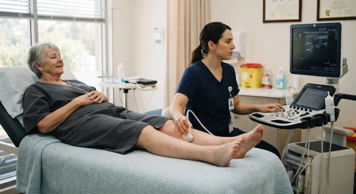
A sonographer applies gel to the skin and moves a transducer over the area being examined. B-mode imaging shows the anatomy, while Doppler measures the direction and speed of blood flow. Blood pressure cuffs may be used for arterial studies, and the sonographer may gently compress veins or ask you to change position or breathe in particular ways. Most examinations are painless, although pressure over a tender area can cause brief discomfort.
Results, recovery and follow-up
No recovery period is needed, and normal activities resume immediately. The vascular surgeon interprets the findings alongside your symptoms and examination, then discusses whether treatment, further imaging or scheduled surveillance is appropriate. Not every narrowing or refluxing vein needs treatment; the scan informs the decision rather than making it.
Surveillance
Repeat ultrasound can monitor aneurysms, carotid disease, stents, bypass grafts, fistulas and treated veins. Scheduled surveillance helps detect changes before symptoms develop.
Risks and limitations
Diagnostic ultrasound is non-invasive and has no known radiation risk, so this section describes limitations rather than complications:
- Image quality can be reduced by body habitus, bowel gas, dressings or heavily calcified vessels
- Some regions, such as the central chest and pelvic veins, are harder to see and may need CT, MR or catheter imaging
- Probe pressure over a tender area can be briefly uncomfortable
- Results are operator-dependent and are always interpreted with the clinical picture
Alternatives and practical considerations
Vascular ultrasound is often the first investigation because it is non-invasive and provides real-time information about blood flow, narrowing, reflux and obstruction. CT, MR or catheter angiography may still be required when vessels are difficult to visualise, when disease extends beyond the ultrasound field or when a procedure is being planned.
The usefulness of the test depends on matching the examination to the clinical question. A venous reflux study, DVT scan, carotid duplex, arterial study and graft surveillance scan use different protocols. Patients should understand what area is being assessed, whether fasting or other preparation is required, and whether the result will be reviewed during a specialist consultation.
Ultrasound findings should not be interpreted in isolation. A narrowing, refluxing vein or anatomical variant may not explain the symptoms, while significant disease can occasionally be underestimated because of technical limitations. The final plan therefore combines the scan with the history, examination and any previous imaging. Repeat studies are often valuable because they show change over time rather than a single measurement.
Vascular ultrasound at Yarra Vascular Surgeons
Yarra Vascular Surgeons integrates ultrasound findings directly with specialist clinical assessment, so each investigation answers a defined question and feeds straight into surveillance or treatment planning without fragmented care.
Arrange a vascular assessment
Vascular ultrasound provides real-time information about flow, narrowing, reflux, thrombosis, aneurysms, grafts and stents without radiation. Ask your GP for a referral, or contact our rooms to arrange a scan with specialist review.